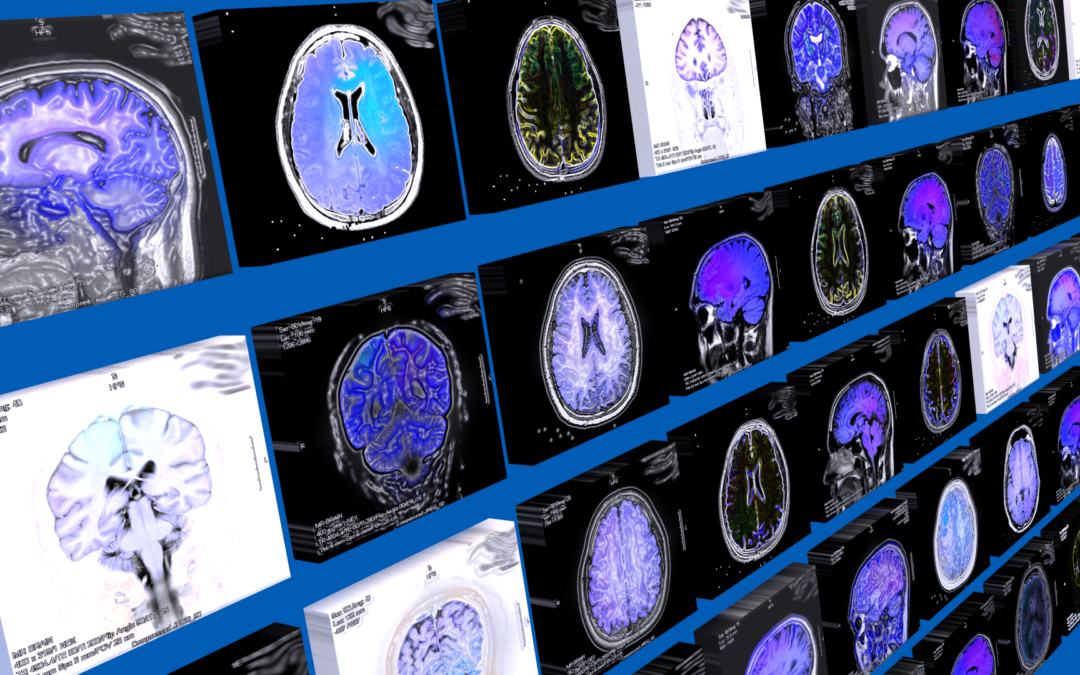
We very often are asked to evaluate individuals who are told they have chronic ischemic white matter changes – punctate lesions – white matter hyperintensities – white matter changes – chronic changes – age related changes – The descriptive list is long as it will reference mostly the same finding on the MRI scan. The individual struggles with the meaning of what the MRI shows because often they do not have the medical conditions that are currently linked to this MRI finding (Example: they do not have diabetes, hypertension, tobacco use, and more). Keeping it simple – what you see is scar. If they do not grow in size or number then the condition is properly treated or has run its local course. If over time there are an increase in number of the lesions then you need to be sure and complete an accurate medical evaluation that will cover all of the main reasons for these lesions. Here we focus a bit on the most important aspect of a hypercoaguable state, in part by showing how complex the physiology truly is. A perturbation of the flow and clotting of blood that ends in an unneeded clot. Keep in mind that while concentration on even the smallest area of damage (10 micron) is important – the radiology team usually will only be able to see areas of damage that are 1000 microns in size. Often even these are too small for them to make specific comments on and the size ends up being more in the range of 5000 microns before it is called a lesion. For the MRI findings there are additional descriptions found in radiology reports and one finds that they are mostly describing the same process. Some additional examples are white matter disease, leukoaraiosis, age related white matter changes, white matter hyperintensities, lacunar infarcts, chronic microvascular changes, chronic microvascular ischemic changes, small vessel disease, periventricular white matter changes – All are terms used to describe that the radiology team can see damage on the brain MRI.


This is one of the main problems that I have seen across many forums – and here it is explained. As someone who comes to this site it was great when I reached out to them on their facebook page and got some additional information! Good work!
What else can be done?